Gaming technology is allowing cancer researchers to imagine – and interact with – cells in a mind-blowing new way.
In a fantastic voyage to the scale of a single cell, scientists can now observe the virtual interaction of therapeutic drugs as they penetrate the very membranes of those cells.
“As well as the obvious educational role this can play, it has the potential to allow cancer researchers to see their data in a new way, which can help them design better nanotherapies,” said John McGhee? at the University of NSW.
Dr McGhee is director of the 3D Visualisation Aesthetics Lab at UNSW’s school of art and design in Paddington. He has harnessed the data from a high-resolution scan of a breast cancer cell to construct a virtual world using technology straight from the gaming industry. He took the data from a University of Queensland scan and built it into a 3D mesh where designers in his lab added colour, lighting, texture and animation.
“It creates a virtual reality world that allows you to be fully immersed in the cell’s architecture,” he said.
It is the first time that data from an actual cell has been used to develop such an interactive virtual reality model.
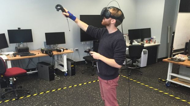
Wearing a VR headset, headphones and holding two hand controls, it is possible to walk across the surface of the cell, as if you have been reduced to the height of 40 one-billionths of a metre.
A flick of a switch allows you to teleport across the cell’s surface to witness a caveolae event, where the cell wall opens to absorb a nanoparticle. Another flick opens a virtual gate that teleports you inside the cell, where you can put your head inside mitochondria and endosomes.

?Different interactive furniture on the cell explain the role of endosomes, or the process of a caveolae event.
?It is a journey like no other that needs to be experienced to fully appreciate its wonder.

Angus Johnston, from Monash University, worked with Dr McGhee to develop the model. His research focuses on the design of therapeutic drugs at the nanoscale, which is a billionth of a metre.
“This technology allows you to see things at the size of the molecule you are trying to deliver,” Dr Johnston said.

The shape, size and chemistry of chemotherapy drugs are all important when designing new therapies to combat cancer. Dr Johnston’s research looks at how drugs can penetrate cell walls and then enter the endosomes of cells before releasing their chemicals.
One way for drugs to enter a cell is diffusion through the cell wall. But this does not allow for targeted delivery to the endosomes, which are transport mechanisms into a cell.

“Visualising the scale of this process will help design the drugs of the future,” he said.
Kara Perrow is research fellow and group leader of the Targeted Cancer Therapeutics Laboratory at the University of Wollongong. She is not connected with the design of the technology.

“It sounds like a great tool for designing drug therapies,” Dr Perrow said. “Seeing modes of entry to cells is very important in the design of targeted nanotherapies. Size has a huge impact on the mechanism of drug uptake.”
Dr Johnston said the model should allow claims about some drugs to be debunked. When some chemical absorption processes are unclear, then imprecise claims about the process can be made. “Looking at the scale of the drug’s interaction will mean we have a better understanding,” he said.

Another cancer researcher unconnected to the technology, UNSW senior lecturer in medicine, Darren Saunders, said spatial and temporal understanding of drug delivery is critical in its design. He was optimistic about its application.
“Looking at a picture of what’s under water is very different to getting into scuba gear and exploring the environment,” Dr Saunders said.
Back at the design lab in Paddington, Dr McGhee said he hopes that the technology will grab the imagination of the next generation of researchers. “The educational opportunities are enormous,” he said. “I hope that this gives high school students, undergrads – as well as researchers – a much better understanding of how the cell functions.”
He said the next step was to include motion data into the model so you can experience the dynamic nature of the cell.
“We are stockpiling terabytes of data about cells that never see the light of day,” Dr McGhee said. “This technology will allow virtual access in every school and museum. It should encourage the best of the best into biology research.”
Dr McGhee’s and Dr Johnston’s work will appear tonight on ABC TV’s Catalyst program.
