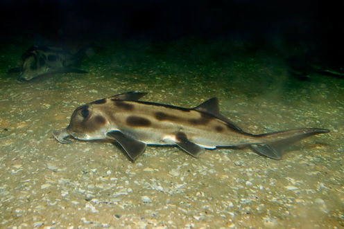
A typical elephant shark from the Melbourne Aquarium. Wikimedia/Fir0002/Flagstaffotos, CC BY-NC
In Greek mythology, looking directly into Medusa’s eyes would turn a person to stone. Yet a similar fate afflicts a very small portion of the population today.
Patients with Fibrodysplasia Ossificans Progressiva, or FOP for short, have their soft tissues turn to bone.
Another abnormality that afflicts humans is Klippel-Feil syndrome, where the individual vertebrae of the neck become fused.
There is no known cure for these diseases at the moment, only treatment, which is more management of the symptoms and prevention of further damage. The gene mutations have been identified but gene therapy and drug therapy have been of very limited help for the conditions.
But in living sharks and rays, and in some fossil armoured fish called placoderms, having a neck encased in bone is the normal condition.
Understanding how this fused neck forms under normal conditions could be the first step in helping us understand how neck development can go wrong when affected by disease.
Rise of the mutants
This is the concept behind evolutionary mutants, where part of an animal’s body mimics what we see in a human disease. Animal models are commonly used to understand disease. But what evolutionary mutants offer that traditional animal models don’t is an insight into how a disease morphology can be formed in a healthy individual.
This is quite different from re-creating the disease in an animal, where we might be getting the same effect (phenotype) but might not be looking at exactly the same underlying causes (genotype).
Elephant sharks, rays and skates are such evolutionary mutants and they are now becoming available for study.
Not only have these animals been around for a long time (more than 400 million years) but they are becoming new model organisms for evolutionary studies.
Elephant sharks, for example, have the slowest evolving genome of any vertebrate. They also have fused vertebrae in the neck region supporting a long fin spine for protection from predators.
Placoderms lived in Australia’s tropical reefs around 375 million years ago and were the top predators of their days with bony armour over their head and thorax. They were the first vertebrate animals with jaws and represent the most basal lineage that we belong to.
Some placoderms also have fused vertebrae in their necks similar to modern elephant sharks. Despite being fossils, they can provide a surprisingly high level of data on how this fusion occurred for comparison to living animals.
The fused neck study
We investigated how this fused neck, called the synarcual, developed in elephant sharks and placoderms, with the results published last month in PLoS One.
We used a growth series of elephant sharks collected from eggs laid in captivity on the Mornington Peninsula, Victoria, and stained them to reveal details of cartilage and muscle development.
We found that neck vertebrae in the elephant shark developed normally, and only later became fused into the synarcual after emerging from their egg capsule.
This is in contrast to the hypothesis that a fused neck forms because individual vertebrae fail to form in early development.
As for the placoderms, we used high-resolution synchrotron scanning at the Australian Synchrotron, Melbourne, to study the development of the synarcual. Placoderms deposit thin bone on their cartilaginous skeletons, which freezes the skeleton at particular growth stages, allowing us to interpret how the synarcual forms.
The individual vertebrae composing the synarcual of the placoderm can still be seen in the synchrotron scan and we found that, as in the elephant shark, the vertebrae that compose the synarcual form individually and normally and only later fuse together to form the synarcual.
Skates and rays appear to show a similar pattern, suggesting that this may be a primitive condition for jawed vertebrate animals as a whole.
The way the synarcual develops in placoderms and sharks is similar to FOP where patients are born with a normal skeleton, with fusion occurring later in life.
Sharks have a kind of hard cartilage instead of true bone, and we don’t fully understand yet if the vertebral fusion is due to over-development of vertebrae, or if the soft tissue between the vertebrae becomes transformed into cartilage, resulting in fusion.
These sorts of transformations of the spine have been observed in farmed salmon and exciting new research is beginning to unravel the genes involved in these transformations. Our goal is to do the same for elephant sharks, rays and skates.
So we are coming closer to understanding how a fused neck develops normally or under stressful conditions (as is the case for farmed salmon) in a range of vertebrates at the base of our ancestry.
The next step is to look further into the gene networks that are responsible for this fusion in our readily accessible evolutionary mutants.
In this way, we might have the key to averting Medusa’s eyes by applying this knowledge to diseases of the human spine.
Catherine Boisvert receives funding from the Australian Research Council.
Kate Trinajstic receives funding from Australian Research Council.
Zerina Johanson receives funding from Australian Research Council
