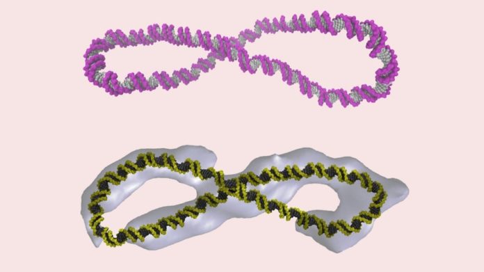
‘Supercoiled’ DNA is more complex than the iconic double helix

DNA doesn’t just coil in the iconic double helix immortalized in every high school biology textbook. It also loops into a menagerie of fantastical shapes, new research finds.
By revealing the hidden shape of DNA, the new insights could provide a more detailed look at the workings of drugs such as chemotherapeutic agents, which interact with DNA.
“This is because the action of drug molecules relies on them recognizing a specific molecular shape — much like a key fits a particular lock,” said study co-author Sarah Harris, a physicist at the University of Leeds in England.
Building blocks of life
After molecular biologists James Watson and Francis Crick first published a paper on the structure of DNA in 1953, the double helix became the iconic symbol of the code of life.
But that picture is actually just a tiny part of nucleic acid structure, researchers now say.
“When Watson and Crick described the DNA double helix, they were looking at a tiny part of a real genome, only about one turn of the double helix. This is about 12 DNA base pairs, which are the building blocks of DNA that form the rungs of the helical ladder,” Harris said.
But DNA is made of about 3 billion base pairs, and all 3.3 feet (1 meter) of this genetic information must fit into the nucleus of a cell, which measures just 10 micrometers across. (For comparison, the average width of a single strand of human hair is 70 micrometers.) To squeeze into such tight quarters, the DNA must be precisely, and tightly, coiled.
Fantastical shapes
To understand this process, the researchers recreated DNA molecules in the lab. Because linear strands of DNA don’t coil, the team painstakingly coiled and uncoiled a helix turn by turn, using short circular snippets of DNA made up of thousands of base pairs.
“Even this relatively modest increase in size reveals a whole new richness in the behavior of the DNA molecule,” Harris said.
The team discovered a panoply of bizarre shapes.
“Some of the circles had sharp bends, some were figure eights, and others looked like handcuffs or racquets or even sewing needles. Some looked like rods because they were so coiled,” the study’s lead author, Rossitza Irobalieva, a biochemist at the Baylor College of Medicine in Houston, said in a statement.
To make sure that this supercoiled DNA actually shows up in the body, the team inserted an enzyme called human topoisomerase II alpha. Just like in the human body, the enzyme relaxed the twist in even the most tightly coiled DNA. This suggests the strangely shaped structures created in the lab mimic the much longer strands of DNA found in the cell nucleus, the researchers reported Oct. 12 in the journal Nature Communications.
Afterward, the team froze the DNA samples and used a special form of microscopy to capture the first-ever images of these fantastical shapes. To get a better look, and to understand how these loops of genetic code act in real-time, the team created computer simulations that revealed the supercoiled loops wriggling over time.
Typically, the DNA helix is formed when complementary base pairs — such as the nucleotide adenine and its partner guanine — bind together, forming a bridge across the helix. But the new simulation revealed that these base-pair bridges peel apart both when the helix is unraveled, and when it is very tightly wound.
The team speculates that base-pair separation in supercoiled DNA allows it to hinge sharply, which could help it cram into the tiny space of a cell’s nucleus.
- ‘Breakthrough’: Ron Howard Talks Science And Tech Innovation | Nat Geo Show Clip
- Human Cyborgs Come to Life in Nat Geo’s ‘Breakthrough’
- Nobel Prize in Literature: 1901-Present
- Myth Busted: Conspiracy Theorists Do Believe Stuff ‘Just Happens’
This article originally published at LiveScience here

