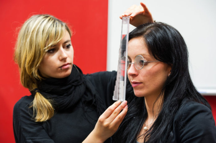
Impaired vision is one of the most common consequences of a stroke. In rare cases, patients may even lose their ability to perceive depth. Such patients see the world around them as flat, like a two-dimensional picture. This makes it impossible for them to judge distances accurately — a skill they need, for instance, when reaching for a cup or when a car is approaching them on the street. A patient with this particular type of visual dysfunction has recently been studied in detail by the research team at Saarland University led by Professor Georg Kerkhoff and Anna-Katharina Schaadt in collaboration with colleagues at the Charité university hospital in Berlin. The team has developed the first effective treatment regime and have identified the area of the brain that, when damaged, may cause loss of binocular depth perception. The results of the study have been published in the academic journal Neuropsychologia.
Strokes can result in a wide variety of visual impairments. ‘A patient may, for example, be blind on one side so that he fails to perceive obstacles or people on that side or have problems when reading,’ explains Georg Kerkhoff, Professor of Clinical Neuropsychology at Saarland University and head of the Neuropsychological Outpatient Service. In some cases, however, the consequences are even more serious. Recently, the team around Kerkhoff and Schaadt collaborated with Professor of Neurology Dr. Stephan Brandt and his colleague Dr. Antje Kraft, both at the Berlin Charité, in treating and supervising a patient who had lost his stereoscopic visual perception as a result of a stroke. Although the patient was able to perceive all the details of his surroundings, he was not able to assess distances with any accuracy. ‘Everything for him was flat, like on a painting,’ explains Anna-Katharina Schaadt, a doctoral research student who is supervised by Kerkhoff and is the study’s lead author. ‘He moved as if in slow-motion and was very uncertain about how far away a coffee cup was on a table or how quickly a car was approaching.’ Like a blind person, he used a long cane to find his way around.
Kerkhoff and Schaadt’s team at the Neuropsychological Outpatient Service on the Saarbrücken campus began by looking for the cause of the patient’s visual impairment.
‘We discovered that the patient was unable to converge the visual impressions from each eye into a single overall image,’ says Schaadt. In healthy individuals, this process is known technically as ‘binocular fusion’ and is important for three-dimensional vision.
Once the diagnosis had been made, the team of neuropsychologists provided a three-week block of therapy during which the patient undertook daily training to improve his visual perception of depth. Three different training methods were employed. Special visual training equipment (prisms, vergence trainer and cheiroscope) were used to present the patient with two images with a slight lateral offset between them. By using what are known as convergent eye movements, the patient attempts to fuse the two images into a single image. This involves directing the eyes inward towards the nose while always keeping the images in the field of view. With time, the two separate images fuse to form a single image that exhibits stereoscopic depth, i.e. the patient has re-established binocular single vision. ‘It was as if a switch had been thrown; the patient was suddenly able to perceive spatial depth again, judge distances correctly and reach out and hold objects with confidence’, describes Schaadt. The patient has now returned to work as a lawyer. At a follow-up examination a year later, the patient still exhibited good stereoscopic depth perception, and can therefore be considered to be permanently cured according to Professor Kerkhoff.
The procedure could be used in future by therapists to help treat other stroke patients suffering from this extreme form of visual impairment. The results of the study are also of interest to researchers working in the field, as Professor Brandt explains: ‘The results illustrate the very specific functional organization of our brains. Damage to the areas known as V6 and V6A in the parietal lobe is associated with impaired three-dimensional visual perception. This area of the brain has been studied in primates. However, further research is required to understand its function in humans.’
Story Source:
The above story is based on materials provided by University Saarland. Note: Materials may be edited for content and length.
Journal Reference:
- Anna-Katharina Schaadt, Stephan A. Brandt, Antje Kraft, Georg Kerkhoff. Holmes and Horrax (1919) revisited: Impaired binocular fusion as a cause of “flat vision” after right parietal brain damage – A case study. Neuropsychologia, 2015; 69: 31 DOI: 10.1016/j.neuropsychologia.2015.01.029
