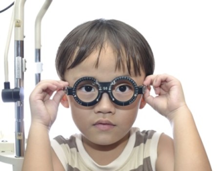
Picture a toddler getting his first eye exam. He’s seated in a strange room, with strange instruments and strange bright lights. He can’t sit still or open his eyes long enough for that diagnostic poof of air — especially if he has trouble seeing anyway, as children with achromatopsia do.
But according to research from the Baylor Visual Function Testing Center, future little ones might not have to squirm in their seats during routine eye exams. The research, which was published in JAMA Ophthalmology, explores a new non-invasive technology that’s kind of like a handheld CT scanner for the eye.
The technology, known as spectral-domain optical coherence tomographic imaging (SD-OCT), helps pediatric ophthalmologists detect achromatopsia by studying retina thickness. It can scan the structure of the eye from a distance, without getting too close to the young patient.
That non-invasive approach is a step up from previous methods, when specialists diagnosed based on age, family history and the standard eye exam procedure (air poof included).
Also known as “day blindness,” achromatopsia is a rare condition that causes bad vision in daylight, color blindness and shaking eyes. It affects one in 40,000 U.S. children and tends to run in families. Worst of all, it’s not easy to predict in young children because the current diagnostic tools were made for grown-ups.
“It has been very difficult to understand the retinal structure of children with achromatopsia because young children are known to be uncooperative during eye examinations designed for the adults,” said Yuquan Wen, PhD, scientific director of the Baylor Visual Function Testing Center. He, along with researchers at the Casey Eye Institute of Oregon Health & Science University, helped develop the study.
As part of the research, investigators studied 18 patients, each of them about 4 years old. Half of the participants suffered from achromatopsia and the other half (control) had normal visual function. By using the SD-OCT, researchers produced 3D high-definition imaging of the kids’ retinas, which is the back part of the eye responsible for creating visual pictures. In many ways, it’s like the film in a camera.
Through those images, they found that the achromatopsia patients had significantly thinner-than-normal retinas, as much as 17 percent thinner than the control participants. The findings imply the importance of studying a child’s retinal thickness when looking for achromatopsia.
Researchers also noted that, in young children, those retinal qualities seemed milder than older patients with the same achromatopsia diagnosis. This could mean a possible therapeutic window to help patients while they’re still young.
“We think that retinal thickness measurement is a more reliable predictor than age alone or genotype alone,” Dr. Wen said. “With the knowledge of retinal thickness in young children with achromaptosia, smarter clinical studies could be designed and monitored based on real structural changes of the retina in conjunction with the visual function change.”
As those new studies take shape, they’ll likely include a form of gene therapy that involves special therapies to make up for the non-functioning genes the patients were born with. Gene therapy has emerged in several clinical trials for blinding eye diseases and likely will continue to do so well into the future.
Before the availability of the handheld SD-OCT, pediatric ophthalmologists had only simple tools and instruments (all of them designed for adults) to detect achromatopsia in children. But based on these findings, the handheld SD-OCT could join those standard tools very soon — as well as be useful in pediatric eye exams in general, Dr. Wen said.
And the squirming kids who endure those eye exams? Like the first-time toddler, things won’t be so tough for them.
Story Source:
The above story is based on materials provided by Baylor Scott & White Health. Note: Materials may be edited for content and length.
Journal Reference:
- Paul Yang, Keith V. Michaels, Robert J. Courtney, Yuquan Wen, Daniel A. Greninger, Leah Reznick, Daniel J. Karr, Lorri B. Wilson, Richard G. Weleber, Mark E. Pennesi. Retinal Morphology of Patients With Achromatopsia During Early Childhood. JAMA Ophthalmology, 2014; 132 (7): 823 DOI: 10.1001/jamaophthalmol.2014.685
