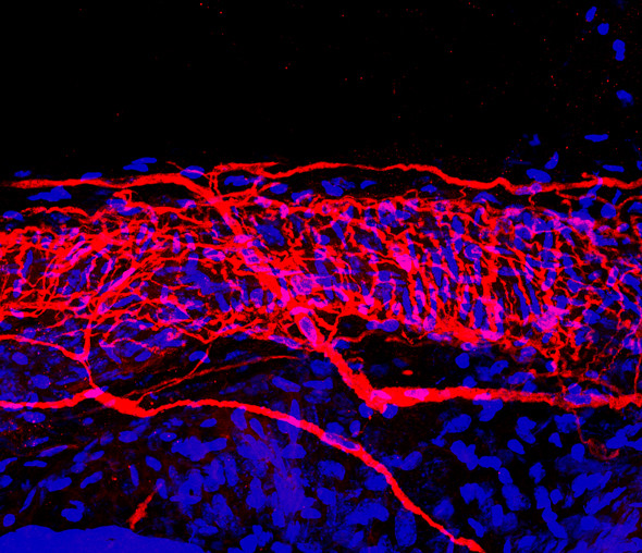
Scientists can now explore nerves in mice in much greater detail than ever before, thanks to an approach developed by scientists at the European Molecular Biology Laboratory (EMBL) in Monterotondo, Italy. The work, published online today in Nature Methods, enables researchers to easily use artificial tags, broadening the range of what they can study and vastly increasing image resolution.
“Already we’ve been able to see things that we couldn’t see before,” says Paul Heppenstall from EMBL, who led the research. “Structures such as nerves arranged around a hair on the skin; we can now see them under the microscope, just as they were presumed to be.”
The technique, called SNAP-tagging, had been used for about a decade in studies using cell cultures — cells grown in a lab dish — but Heppenstall’s group is the first to apply it to neurons in living mice. It allows researchers to use virtually any labels they want, making it easier to overcome the challenges that often come with studying complex tissues and animals. To study nerves in the skin, for instance, Heppenstall’s lab can employ artificial dies that are small enough to cross the barrier posed by the skin itself, and stand out better from the skin’s natural fluorescence. And because these are artificial, custom-made tags, they can be designed to do more than just highlight particular structures. Scientists can produce tags that destroy certain structures or cells, for instance.
SNAP-tagging relies on a small protein that binds to a specific small chemical structure — and once bound, it won’t let go. The EMBL scientists genetically engineered mice so that their cells would produce that SNAP protein. They then used fluorescent probes that contain the small chemical that SNAP binds to, and injected them into the mice. SNAP acts like an anchor, glueing the tags in place for researchers to follow under the microscope.
Ultimately, Heppenstall aims to employ this approach to record activity in individual neurons. For instance, he’d like to mechanically stimulate the skin, or change its temperature, and watch that information flow through the nerve, to the next nerve, tracking it throughout the whole network. In principle, he speculates, you could do this in a whole brain. It would be like a taking a scan and zooming in to see what’s happening inside each nerve cell.
Story Source:
The above story is based on materials provided by European Molecular Biology Laboratory (EMBL). Note: Materials may be edited for content and length.
Journal Reference:
- Guoying Yang, Fernanda de Castro Reis, Mayya Sundukova, Sofia Pimpinella, Antonino Asaro, Laura Castaldi, Laura Batti, Daniel Bilbao, Luc Reymond, Kai Johnsson, Paul A Heppenstall. Genetic targeting of chemical indicators in vivo. Nature Methods, 2014; DOI: 10.1038/nmeth.3207
