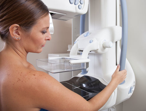
We’ve known for some time that women with dense breasts are at higher risk of breast cancers that are difficult to detect by mammography. To address this problem, 19 US states have introduced laws requiring health providers to notify women whose mammograms show they have “dense breasts”.
These women are encouraged to discuss this finding with their doctor and, if necessary, have follow-up ultrasounds or magnetic resonance imaging scans to look for possible breast cancers.
But new US modelling testing the impact of routine follow-up ultrasounds for women with dense breasts found the process would be costly, lead to unnecessary anxiety and deliver little benefit.
While we’re yet to find a better way to detect breast cancers in women with dense breasts, directing all women with dense breasts to have follow-up imaging might be an unwarranted and costly public health strategy.
Why does density matter?
“Breast density” does not refer to how hard a woman’s breasts feel. It refers to the white or bright areas of a mammogram and is more appropriately referred to as “mammographic density”.
Mammographic density is important for two reasons. First, and in the context of the US laws, the brighter areas on the mammogram can cover up existing breast tumours. This is referred to as “masking”.
Australian researchers have been studying the masking phenomenon for many years and have found the use of hormone therapy could increase the problem for some women. Therefore an “all clear” from a mammogram can be a “false negative”.
Second, for women of the same age and body mass index, those with higher mammographic density are at greater risk of a future breast cancer. The one-quarter of women in the highest-density category are almost three times more likely to develop breast cancer than the one-quarter of women in the lowest-density category.
The predictive power is not dramatic on a personal level, as it is for having a mutation in a breast cancer susceptibility gene such as BRCA1 or BRCA2. But because women vary so much in their mammographic density, on a population basis it is among the strongest risk factors for breast cancer.
Genetic link
In collaboration with BreastScreen services across Australia and colleagues in Canada, my research team undertook a large twin study and found genetic factors play an important role in breast density variation.
From this, we predicted that mammographic density explains about 10% of why breast cancer runs in families. New genetic studies are confirming this prediction.
We also identified the first gene that influences both mammographic density and breast cancer, called LSP1. A large international collaboration led by Associate Professor Jennifer Stone at the University of Western Australia has found at least ten more such genes. This research will soon be published.
We have also found that mammographic density measures that predict breast cancer risk “track” through life. This means mammographic density in early adulthood predicts mammographic density in mid-life, and hence breast cancer risk.
Towards better detection
We’re yet to find an alternative way to better detect breast cancer in women with dense breasts. But international researchers are working to:
-
develop automated measures using digital mammograms that best predict breast cancer risk
-
develop optimal screening protocols (ages and times between mammograms) for women, depending on their mammographic density
-
determine the most cost-effective alternative screening strategies for women with mammographically dense breasts
-
implement these strategies in breast screening services, including those in developing countries in which breast cancer is becoming an increasingly important disease.
While we’re making substantial progress in the first two areas, there is a long way to go for the last two.
Another area of research is to find out what can be done to lower a woman’s mammographic density and hence, it is hoped, her risk of breast cancer.
We know, for instance, that child birth gives a small but long-term reduction in breast cancer risk. During pregnancy, the breasts becomes mammographically dense, but after lactation the density reduces by an average of 7% when compared with the pre-pregnancy density.
There is also some evidence that tamoxifen, a drug given to reduce breast cancer risk, especially for women who have had a breast cancer, might also reduce mammographic density.
Mammographic density also appears to explain why having a high body mass index in late adolescence is associated with being at lower risk of breast cancer.
So dense breasts are not a new phenomenon. Australian researchers and BreastScreen have been working hard for nearly two decades to understand and, most importantly, work out how to use this concept to reduce deaths from breast cancer.
With the increasing availability of digital mammography, breast density is more easily measured, and researchers continue to search for better ways to detect breast cancer in women with dense breasts.
The challenge now is to use mammographic density to help make breast screening more effective in preventing deaths from breast cancer, at lower financial cost and with fewer of the negative impacts inherent in all population-wide screening programs.
John Hopper receives funding from the National Breast Cancer Foundation, Cancer Council Australia, Cancer Australia, National Institutes of Health in the US, VicHealth.
