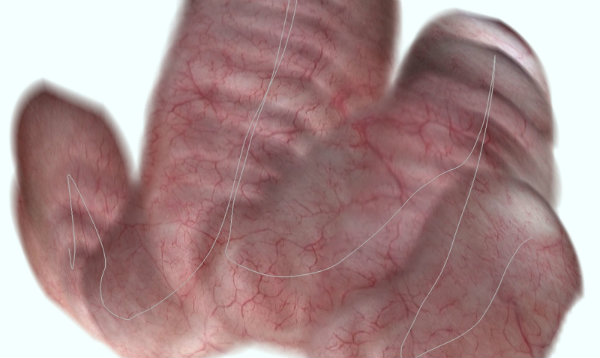
The software provides a panoramic perspective made from images of the bladder‘s inside wall that were taken endoscopically. As an option, it can also depict which pathways the physician has taken using the endoscope (grey line). © Fraunhofer IIS
Physicians performing cystoscopies get a tunnel-like view of the insides, being able to only see a focused image a short distance at a time. Reviewing the entire path of the cystoscope in one image is now being made possible by researchers at the Fraunhofer Institute for Integrated Circuits in Erlangen, Germany.
The Endorama software they developed automatically stitches together the images captured as the cystoscope is edvanced through the urethra and bladder to its final destination. This is done nearly in real-time, allowing the physician to quickly see whether the entire path has been captured and properly reviewed. Moreover, while previously the physician had to draw a mental picture of what is being examined from visualizing different spots independently, the new software provides a more intuitive look that lets the clinician focus on performing the procedure rather than overcoming the endoscope’s limitations.
From Fraunhofer:
The software takes the image that the camera currently records and in each case displays it at the center of the screen. If the computer panorama displays a ”hole” at some point, then the physician knows that he has not yet examined the bladder wall at that point. ‘Endorama’ also provides advantages even when it comes to documentation and records: Instead of a single image or a video file, the physician can stitch together the panoramic image into the patient files, because this image depicts the entire investigative findings and additionally documents that the bladder was studied seamlessly.
In order to prepare a panorama like this, the camera on the endoscope acquires about 25 pictures a second, which overlap each other. The software searches for distinctive features in the images and based on these structures, compiles them into a complete overall picture. Whereas panoramic images are common in conventional photography, and can be taken with the right app on a smart phone, for example, they also harbor a number of challenges when it comes to endoscopy imaging: Optically, these images as a rule are sharply distorted, feature low resolution and even the image contrast is relatively minimal due to the irregular lighting. In addition, the structures in the bladder are weakly expressed; therefore, it is important to find distinctive points, on the basis of which the overlapping images can be compiled. ‘Endorama’ makes all this possible: In a first step, the software corrects the optical distortions and balances out the shadows that arise due to the non-homogenous lighting. Various computer processes assemble the images: while one process searches for designated image features, such as vascular structures on the bladder wall, another is fitting the images together in an organized manner. In doing so, these processes also take into account the complex geometry of the bladder.
‘Endorama’ has already passed initial tests successfully: The researchers reviewed the software initially using a phantom configuration; a ten-centimeter plastic ball on whose inner side the vascular structure of the bladder was mapped. The scientists also assembled video sequences which were obtained from standard bladder examinations into panoramas.
Press release: Endoscopy with panoramic view…
(hat tip: Gizmodo)
