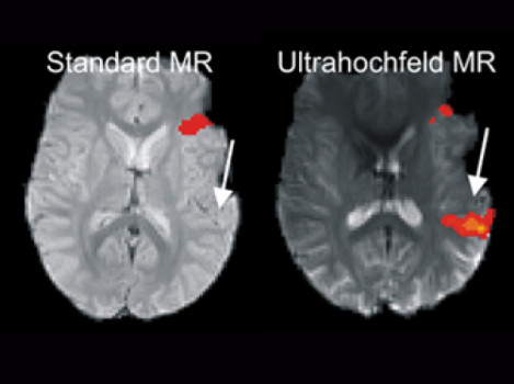
In a new investigation by the University Department of Neurology, it has been possible for the first time to demonstrate that the areas of the brain that are important for understanding language can be pinpointed much more accurately using ultra-high-field MRI (7 Tesla) than with conventional clinical MRI scanners. This helps to protect these areas more effectively during brain surgery and avoid accidentally damaging it.
Before brain surgery, it is important to precisely understand the areas of the brain required for language in order to avoid injuring them during the procedure. Their position can shift considerably, especially in patients with tumours or brain injuries. The brain’s flexibility also means that language centres can shift to other regions. If the areas responsible for language control and processing are injured during a brain operation, the patient can be left unable to communicate. In order to create a “map” of the language control centres prior to the operation, functional magnetic resonance imaging (fMRI) is used these days.
A multi-centre study from 2013 demonstrated the advantages of fMRI-assisted localisation of the motor centres in the brain. In a new investigation by the working group led by Roland Beisteiner (University Department of Neurology), it has been possible for the first time to demonstrate that the areas of the brain that are important for understanding language can be pinpointed even more accurately using ultra-high-field MRI (7 Tesla) than with conventional clinical MRI scanners. The focus lies on the two most important language centres in the brain known as Wernicke’s area (which controls the understanding of language) and Broca’s area (which controls the motor functions involved with speech).
The brain is scanned for activity while the patient is carrying out speech exercises. This allows the areas required for speech to be localised much more accurately than previously. “Ultra-high-field MR offers much greater sensitivity than classic MRI scanners,” explains Roland Beisteiner, “allowing even very weak signals to be recorded in areas that would otherwise have been missed.”
The work was carried out in cooperation between the University Department of Radiology and Nuclear Medicine and other university departments as well as with support from one of the research clusters at Vienna’s universities (Roland Beisteiner, Tecumseh Fitch) and was published in the highly respected journal Neuroimage.
In Austria, fMRI methods were first set up and established for clinical use by the University Department of Neurology back in 1992. They are funded exclusively through third-party finance and have been constantly developed in cooperation with other university organisations. As a result, the working group has published numerous pioneering papers on the improvement of neurological diagnostics using fMRI and carried out the world’s first study into improving the diagnosis of brain function using ultra-high-field MRI. Today, fMRI is the most important non-invasive technique in Austria for the research and clinical exploration of brain functions.
Story Source:
The above story is based on materials provided by Medical University of Vienna. Note: Materials may be edited for content and length.
Journal Reference:
- A. Geißler, E. Matt, F. Fischmeister, M. Wurnig, B. Dymerska, E. Knosp, M. Feucht, S. Trattnig, E. Auff, W.T. Fitch, S. Robinson, R. Beisteiner. Differential functional benefits of ultra highfield MR systems within the language network. NeuroImage, 2014; 103: 163 DOI: 10.1016/j.neuroimage.2014.09.036
