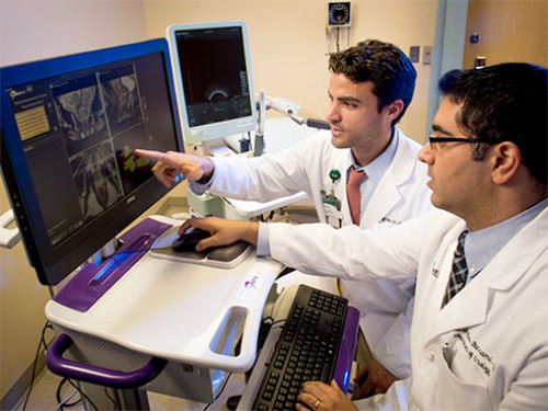
The latest advancement in prostate cancer detection is magnetic resonance imaging and ultrasound fusion-guided biopsy, which offers benefits for both patient and physician.
The only place in the Southeast offering the MRI-US image fusion technique is at the University of Alabama at Birmingham Program for Personalized Prostate Cancer Care.
It is estimated that 2014 will see more than 240,000 new cases of prostate cancer, and more than 29,000 deaths from the disease, according to the National Cancer Institute.
Jeffrey Nix, M.D., along with colleague Soroush Rais-Bahrami, M.D., both assistant professors in the UAB Department of Urology, studied the MRI-US image fusion as fellows at the NCI. Nix and Rais-Bahrami are two of a select few urologists in the United States trained to utilize this technology; together they have five years’ experience using this approach.
Nix and Rais-Bahrami say this new technology offers a “targeted biopsy,” which refers to direct tissue sampling of suspicious areas seen on MRI as opposed to the traditional method of random, systematic sampling that is essentially performed “blindly” in different “ZIP code” regions of the prostate.
“We are utilizing prostate MRI and fusing it with real-time ultrasound for image-guided prostate biopsies; this can detect prostate cancer with high accuracy, and it accurately targets lesions of concern defined by MRI,” Nix said. “This improves overall detection compared to standard biopsy and, more importantly, has the potential to give clinicians and patients a more accurate picture of their true disease burden by allowing improvements in staging.”
Studies of this new technique, Nix says, have shown that it increases the overall cancer detection rate, increases the high-risk cancer detection rate, and improves staging for patients who are considering active surveillance, which is when your doctor closely monitors your low-risk prostate cancer for any changes.
“The technique is expected to be especially helpful in cases of men with a history of negative biopsies who are still suspected of having cancer due to a persistently unexplained elevated prostate-specific antigen level, patients with enlarged prostates and patients being guided toward active surveillance for improved staging,” said Rais-Bahrami.
Rais-Bahrami adds that MRI-US fusion-guided biopsy is a clinic-based procedure that can be performed under local anesthesia; the patient’s experience of this new biopsy versus traditional biopsy without MRI guidance is the same, but with more accurate outcomes based on the targeting approach.
“I have a patient who had five previous biopsy sessions over the past seven years, and he’s had persistently elevated PSA, yet each biopsy came back negative,” Rais-Bahrami said. “When he came to us and had the MRI-US fusion-guided biopsy, we were able to target areas that we identified with our radiologists as areas of concern, and one in fact came back as cancerous. This is probably what’s been there causing his PSA elevation all this time; however, it was hidden to all these biopsy sessions over the past seven years.”
“We’ve been offering this technology at UAB for the last year, and we’ve seen a lot of success,” Nix said. “I have had several patients who were on active surveillance, and the MRI-US fusion biopsy discovered significantly more extensive disease. Those patients were able to go on to treatment and to cancer cure. It turned out some patients had prostate cancer even after they had multiple biopsies that came back negative; this enabled them to make more informed decisions on appropriate treatment.”
“This is the first major advancement in prostate cancer detection in more than 30 years, and it’s a significant improvement,” Nix said.
Story Source:
The above story is based on materials provided by University of Alabama at Birmingham. The original article was written by Nicole Wyatt. Note: Materials may be edited for content and length.
