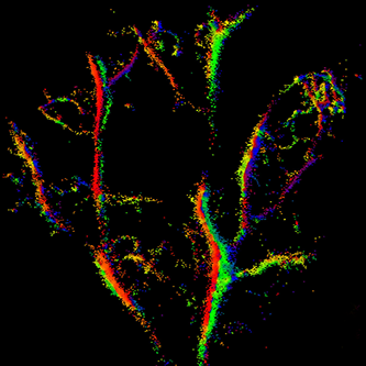
While you can check out the carotid artery using ultrasound probes, imaging much smaller blood vessels that permeate our bodies is impossible using conventional ultrasound due to its inherent diffraction limit. But, just like with light, a bit of trickery and ingenuity can overcome what was previously thought of as a fundamental physical barrier.
Researchers at Kings College London devised a technique during which microbubbles about the size of red blood cells were injected into the ear of a mouse. The bubbles were delivered in a moderate quantity so that there was enough, but not too many of them, so that each would be noticed using ultrasound yet not overwhelm and oversaturate the entire image. Being able to track individual bubbles as they moved through the microvasculature of the mouse ear resulted in a velocity map of the blood network at a resolution only possible with optical microscopes (and dissection).
From the study abstract in IEEE Transactions on Medical Imaging:
Resolution is improved from a measured lateral and axial resolution of 112 μm and 94 μm respectively in original US data, to super-resolved images of microvasculature where vessel features as fine as 19 μm are clearly visualized. Velocity maps clearly distinguish opposing flow direction and separated speed distributions in adjacent vessels, thereby enabling further differentiation between vessels otherwise not spatially separated in the image. This technique overcomes the diffraction limit to provide a non-invasive means of imaging the microvasculature at super-resolution, to depths of many centimeters. In the future, this method could noninvasively image pathological or therapeutic changes in the microvasculature at centimeter depths in vivo.
Study in IEEE Transactions on Medical Imaging: In Vivo Acoustic Super-Resolution and Super-Resolved Velocity Mapping Using Microbubbles…
King’s College: Mapping blood flow with bubbles and ultrasound…
