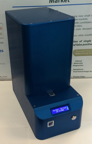
Ebola is no longer a disease that only Dustin Hoffman is paranoid about. People around the world are worried that we might have a pandemic on our hands that modern medical technology may not be able to stop. Yet, researchers around the world aren’t sitting on their hands waiting to be consumed by the disease and the fear it engenders. One major part in being able to control the spread of Ebola is early identification of infected patients. Currently, time consuming and expensive fluorescent label-based techniques are used to spot the Ebola virus in blood samples, often allowing the disease to spread faster than it can be tracked down. Scientists at Boston University have been developing a new photonic device that doesn’t require labeling of samples and may provide quick and accurate diagnosis of Ebola anywhere it is taken.
The device has already been shown to be able to spot individual H1N1 virus particles, but now the researchers have shown that it can identify multiple viruses at the same time, including viruses that were genetically modified to resemble Ebola and Marburg.
Previous techniques generally required a few hours of preparation and processing before results were provided, but the new device demands very little prep and spits out results within about one hour.
The device is a so called “single particle interferometric reflectance imaging sensor (SP-IRIS)” that shines multi-colored light onto a surface coated with anti-bodies that attract specific viruses. The reflection of this light is affected by viruses that are stuck to the coated surface, and the size and type of the virus particle defines how that light appears to a light sensor.
From the study abstract in ACS Nano:
We demonstrate identification of virus particles in complex samples for replication-competent wild-type vesicular stomatitis virus (VSV), defective VSV, and Ebola- and Marburg-pseudotyped VSV with high sensitivity and specificity. Size discrimination of the imaged nanoparticles (virions) allows differentiation between modified viruses having different genome lengths and facilitates a reduction in the counting of nonspecifically bound particles to achieve a limit-of-detection (LOD) of 5 × 10³ pfu/mL for the Ebola and Marburg VSV pseudotypes. We demonstrate the simultaneous detection of multiple viruses in a single sample (composed of serum or whole blood) for screening applications and uncompromised detection capabilities in samples contaminated with high levels of bacteria. By employing affinity-based capture, size discrimination, and a “digital” detection scheme to count single virus particles, we show that a robust and sensitive virus/nanoparticle sensing assay can be established for targets in complex samples. The nanoparticle microscopy system is termed the Single Particle Interferometric Reflectance Imaging Sensor (SP-IRIS) and is capable of high-throughput and rapid sizing of large numbers of biological nanoparticles on an antibody microarray for research and diagnostic applications.
Boston University: Containing Ebola with Nanotechnology…
