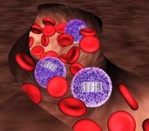
A 7-year-project to develop a barcoding and tracking system for tissue stem cells has revealed previously unrecognized features of normal blood production: New data from Harvard Stem Cell Institute scientists at Boston Children’s Hospital suggests, surprisingly, that the billions of blood cells that we produce each day are made not by blood stem cells, but rather their less pluripotent descendants, called progenitor cells. The researchers hypothesize that blood comes from stable populations of different long-lived progenitor cells that are responsible for giving rise to specific blood cell types, while blood stem cells likely act as essential reserves.
The work, supported by a National Institutes of Health Director’s New Innovator Award and published in Nature, suggests that progenitor cells could potentially be just as valuable as blood stem cells for blood regeneration therapies.
This new research challenges what textbooks have long read: That blood stem cells maintain the day-to-day renewal of blood, a conclusion drawn from their importance in re-establishing blood cell populations after bone marrow transplants — a fact that still remains true. But because of a lack of tools to study how blood forms in a normal context, nobody had been able to track the origin of blood cells without doing a transplant.
Boston Children’s Hospital scientist Fernando Camargo, PhD, and his postdoctoral fellow Jianlong Sun, PhD, addressed this problem with a tool that generates a unique barcode in the DNA of all blood stem cells and their progenitor cells in a mouse. When a tagged cell divides, all of its descendant cells possess the same barcode. This biological inventory system makes it possible to determine the number of stem cells/progenitors being used to make blood and how long they live, as well as answer fundamental questions about where individual blood cells come from.
“There’s never been such a robust experimental method that could allow people to look at lineage relationships between mature cell types in the body without doing transplantation,” Sun said. “One of the major directions we can now go is to revisit the entire blood cell hierarchy and see how the current knowledge holds true when we use this internal labeling system.”
“People have tried using viruses to tag blood cells in the past, but the cells needed to be taken out of the body, infected, and re-transplanted, which raised a number of issues,” said Camargo, who is a member of Children’s Stem Cell Program and an associate professor in Harvard University’s Department of Stem Cell and Regenerative Biology. “I wanted to figure out a way to label blood cells inside of the body, and the best idea I had was to use mobile genetic elements called transposons.”
A transposon is a piece of genetic code that can jump to a random point in DNA when exposed to an enzyme called transposase. Camargo’s approach works using transgenic mice that possess a single fish-derived transposon in all of their blood cells. When one of these mice is exposed to transposase, each of its blood cells’ transposons changes location. The location in the DNA where a transposon moves acts as an individual cell’s barcode, so that if the mouse’s blood is taken a few months later, any cells with the same transposon location can be linked back to its parent cell.
The transposon barcode system took Camargo and Sun seven years to develop, and was one of Camargo’s first projects when he opened his own lab at the Whitehead Institute for Biomedical Research directly out of grad school. Sun joined the project after three years of setbacks, and accomplished an experimental tour de force to reach the conclusions in the Nature paper, which includes data on how many stem cells or progenitor cells contribute to the formation of immune cells in mouse blood.
With the original question of how blood arises in a non-transplant context answered, the researchers are now planning to explore many more applications for their barcode tool.
“We are also tremendously excited to use this tool to barcode and track descendants of different stem cells or progenitor cells for a range of conditions, from aging, to the normal immune response,” Sun said. “We first used this technology for blood analysis, however, this system can also help address basic questions about cell populations in solid tissue. You can imagine being able to look at tumor progression or identify the precise origins of cancer cells that have broken off from a tumor and are now circulating in the blood.”
“I think that not only for the blood field, this can change the way people look at stem cell and progenitor relationships,” Camargo added. “The feedback that we have received from other experts in the field has been fantastic. This can truly be a groundbreaking technology.”
Story Source:
The above story is based on materials provided by Harvard University. Note: Materials may be edited for content and length.
Journal Reference:
- Jianlong Sun, Azucena Ramos, Brad Chapman, Jonathan B. Johnnidis, Linda Le, Yu-Jui Ho, Allon Klein, Oliver Hofmann, Fernando D. Camargo. Clonal dynamics of native haematopoiesis. Nature, 2014; DOI: 10.1038/nature13824
