Medical breakthrough as researchers uncover the mystery of how hematopoietic stem cells form

Australian researchers studying zebrafish have made one of the most significant ever discoveries in stem cell research.
They have uncovered the mystery of how a critical type of stem cell found in blood and bone marrow, and essential to replenishing the body’s supply of blood and immune cells, is formed.
The cells, called hematopoietic stem cells (HSC), are already used in transplants for patients with blood cancers such as leukemia and myeloma.
But HSCs have significant potential to treat a broader range of conditions because they appear to be able to form all kinds of vital cells including muscle, blood vessel and bone.
The problem was scientists had no idea how HSCs formed, making growing them in a lab and using them to treat spinal cord injuries, diabetes and degenerative disorders impossible.
However, a research team led by Professor Peter Currie, from the Australian Regenerative Medicine Institute at Victoria’s Monash University, uncovered a major part of HSC’s development. Understanding how HSCs self-renew to replenish blood cells is considered the holy grail of advancing stem cell research.
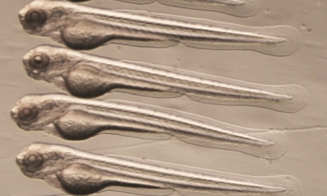
Using high-resolution microscopy, Currie’s team filmed HSCs as they formed inside zebrafish embryos. In playing the film back, they saw a “buddy” cell appeared to help HSCs form.
“It’s a sad fact of life that humans are basically just modified fish, and our genomes are virtually identical to theirs,” Currie said.
“Zebrafish make HSCs in exactly the same way as humans do, but what’s special about these guys is that their embryos and larvae develop free living and not in utero as they do in humans.
“So not only are these larvae free-swimming, but they are also transparent, so we could see every cell in the body forming, including HSCs.”
The researchers were initially studying muscle mutations in the zebrafish. But when playing the film back they noticed that the muscle-deficient zebrafish had several times the normal population of HSCs.
They saw the pre-HSCs required a “buddy” cell, known as endotome cells, to turn into HSCs.
“Endotome cells act like a comfy sofa for pre-HSCs to snuggle into, helping them progress to become fully fledged stem cells,” Currie said.
“Not only did we identify some of the cells and signals required for HSC formation, we also pinpointed the genes required for endotome formation in the first place.
“I’m not an HSC biologist, I’m an muscle cell biologist, so this was a highly serendipitous finding we made because these helper cells are made next to the muscle stem cells we were initially examining.”
He said researchers could now focus on finding the signals present in the endotome cells responsible for HSC formation in the embryo.
“Then we can use them in the lab to make different blood cells on demand for all sorts of blood-related disorders,” he said.
If they could do this, there would also be the potential for genetic defects in cells to be corrected and transplanted back into the body, he said.
Their findings were published in the prestigious international journal Nature on Thursday.
Dr Georgina Hollway, from the Garvan Institute of Medical Research in Sydney, said the work highlighted how molecular processes in the body play a key role in HSC formation.
“We now know that these migratory cells are essential in the formation of HSCs, and we have described some of the molecular processes involved,” Hollway said.
“This information is not the whole solution to creating them in the lab, but it will certainly help.
“It’s difficult to say exactly how close we are, but we have uncovered a vital step in the process.”
